AV Valves and Semilunar Valves: The major distinction between AV valves and semilunar valves lies in their respective functions; an AV valve allows blood to move from atria to ventricles while semilunar valves allow blood from ventricles to emergent arteries of ventricles.
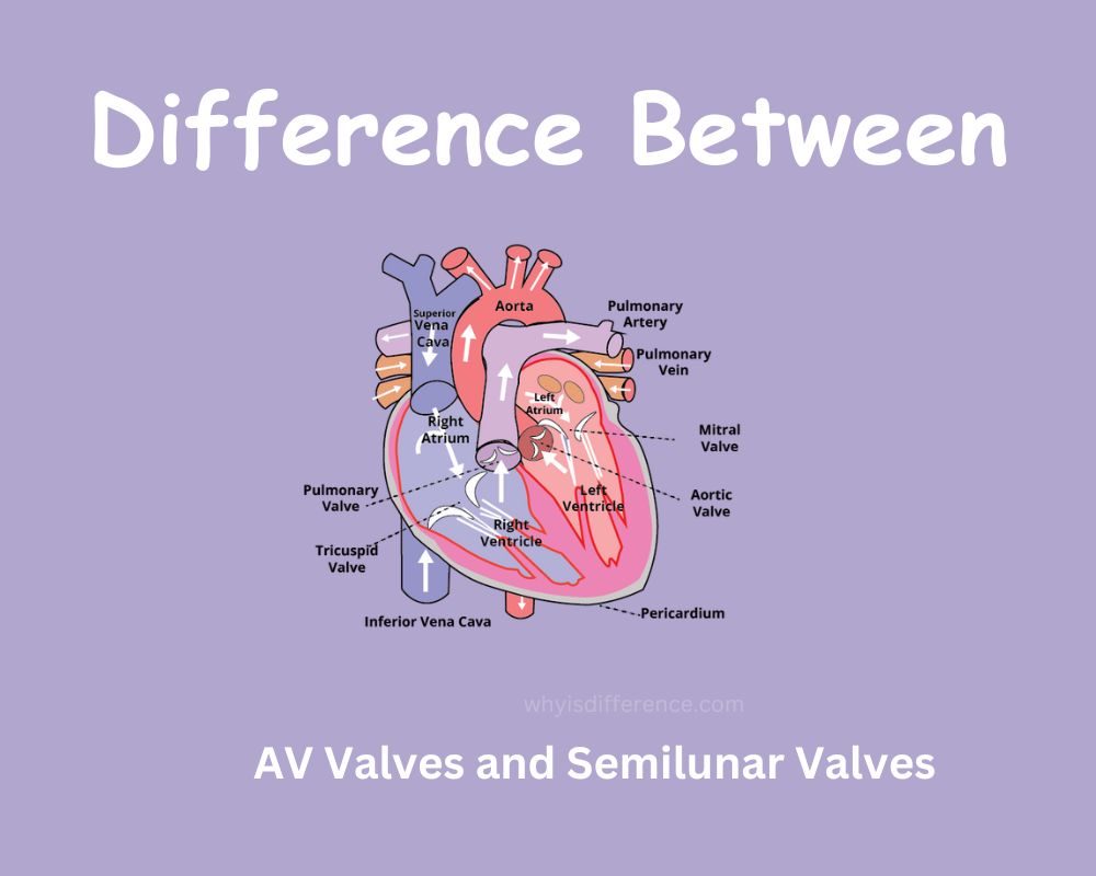
The heart is a muscular organ responsible for pumping blood throughout our bodies via our circulatory system, providing oxygen and other essential nutrients while cleansing away metabolic wastes from them. A human heart contains four chambers two atria, the left ventricle, and the right ventricle. Furthermore, four heart valves enable blood to move in one direction through it the mitral valve and tricuspid valve as well as semilunar valves. When coupled with their respective sides on either side of their respective sides, blood is kept flowing freely throughout your entire system and cleanly rid yourself of metabolic wastes from within.
Overview of the Cardiovascular System
The circulatory system, commonly referred to as the cardiovascular system, is responsible for transporting blood, oxygen, and nutrients throughout the body. Composed of blood vessels, blood, and the heart, it transports vital life blood.
The Heart:
The heart is an organ with muscular structures located within the chest cavity. Its purpose is to deliver oxygenated and deoxygenated body blood into tissues and organs of the body and is made up of four chambers two atria on either side (left/right) as well as two ventricles on each side.
As part of a double pump system, its right side receives blood from body tissues before pumping it to be oxygenated in the lungs while the left side takes in oxygenated blood from these lungs before receiving oxygenated blood for processing before sending it onwards into tissues/organs/body before receiving deoxygenated blood from body tissues/organs/ organs from the body; left side only receiving oxygenated blood from chest cavity/lungs for distribution throughout.
Blood Vessels:
Arteries: Arteries are vessels that carry oxygenated blood from the heart to other parts of the body. Their walls are thick and elastic to help control blood pressure.
Veins: Veins are blood vessels that return deoxygenated body blood back to the heart after traveling through tissues and organs, unlike arteries which carry it forward from these sources. Veins differ in that their walls have thinner walls; additionally, they feature valves to prevent backflow.
Blood: Blood is an essential fluid that supplies oxygen and nutrients throughout the body. Composed of platelets, plasma, red blood cells, and white blood cells–both important parts of immune defense systems and carriers of oxygen–platelets perform blood clotting duties while plasma acts as an envelope for cells and substances contained within it.
The cardiovascular system works hard to deliver oxygen and nutrients to tissues throughout the body while eliminating waste products, creating homeostasis, and supporting the proper functioning of all systems of the body. It is an indispensable system essential to health.
AV Valve (Atrioventricular Valves)
AV Valve (also referred to as Atrioventricular or AV Valve) is located within the heart and regulates blood flow between ventricles and atria, thus ensuring it stays on course and preventing backflow during cardiac cycles.
Definition and Location:
- The AV valves in a human heart are located between its atria (upper chambers) and ventricles (lower chambers), two being present.
- The Mitral Valve lies between the left ventricle and the left atrium of your body.
- Tricuspid Valve This valve lies between the right atrium and the ventricle.
Structure and Composition:
- The AV valves consist of strong fibrous tissue composed of multiple components.
- Valve leaflets/cusps are thin flaps of tissue that open and close in order to either allow blood to flow into or stop it from flowing out of a vessel.
- Supportive valve leaflets help prevent inverting into the atria during ventricular contraction, offering crucial support. In turn, these supports prevent valve leaflets from inverting backward into the atrium when the ventricles contract and vice versa.
- the leaflets of each valve are connected by a fibrous ring known as an annulus that serves to secure it into place. This anchor ensures optimal functioning.
Function:
The primary function of AV valves is to regulate blood flow between the atria and ventricles using various mechanisms:
a. Opening and Closing:
The AV valves are opened and closed by changes in heart chamber pressure during cardiac cycles. For instance, they open when ventricles relax during diastole, allowing blood from the atria to flow into ventricles; when ventricles constrict, however, they close to prevent backflow into the atria.
b. Blood Flow Regulation:
The AV valves ensure unidirectional blood flow by closing when ventricles contract to prevent blood from flowing back into the atria; instead, it’s directed into major arteries such as the aorta or pulmonary artery for distribution throughout the body.
Relationship Between Atria Ventricles and Ventricles:
The AV valves play a pivotal role in coordinating contraction and relaxation between the atria, ventricles, and between them.
a. Atrial Contraction and Aortic Valve Closure:
When the atria contract, the AV valves close to prevent blood from flowing backward into the atria and back into circulation.
b. Ventricular Relaxation and Aortic Valve Opening:
As the ventricles relax, their pressure drops significantly, prompting AV valves to open due to this pressure difference and allow blood from the atria into the ventricles.
Assuring the proper functioning of the AVs is key for efficient blood circulation in the heart. Doing so prevents backflow of blood which could otherwise lead to cardiovascular issues and decreased cardiac performance.
Semilunar Valves
Semilunar valves in the heart regulate blood flow between ventricles, major arteries, and aorta to ensure it flows in an orderly fashion and stops backflow in its cycle. These valves ensure blood travels in the right direction without backflow occurring during cardiac contractions and relaxations.
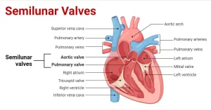
Definition and Location:
Semilunar valves can be found at the base of two major arteries that leave the heart. Two semilunar arteries leave as well.
- a.This valve lies between the left ventricle and aorta (the main artery that transports oxygenated blood around your system), and controls flow into it from both locations.
- b. The pulmonary valve lies between the right ventricle and pulmonary artery and transports deoxygenated blood into the lungs to be oxygenated.
Structure and composition:
Semilunar valves consist of three semilunar cusps or leaflets and derive their name from their crescent-shaped crescent cusps or halves, as well as their thin fibrous tissue leaflets that lack papillary or chordae tendineae structures.
Functions:
Semilunar valves serve to regulate blood flow from ventricles to major arteries by using various mechanisms.
Opening and Closing:
Semilunar heart valves respond to pressure changes within the chambers of the heart. Semilunar valves open when pressure increases in the ventricles (ventricular systole), allowing blood to move from these chambers towards respective arteries; they close again during ventricular diastole when ventricles relax (ventricular diastole).
Semilunar valves help prevent backflow by only allowing one direction of blood flow between ventricles and major arteries, closing when ventricles relax to stop backflow blood from the aorta or pulmonary artery from flowing back into them and maintaining forward blood flow for efficient circulation. This ensures forward flow rather than backflow of blood ensuring efficient circulation by maintaining forward flow of blood from ventricles to major arteries.
Relationship Between Ventricles and Major Arteries:
Semilunar valves play an essential role in the coordination of contraction and relaxation of ventricles and their flow into major arteries.
Semilunar valve opening and ventricular contraction:
As soon as the ventricles contract, their pressure increases beyond that in major arteries; this pressure differential opens semilunar vales to let blood from within the ventricles enter either aorta or the pulmonary artery.
Ventricular relaxation and semilunar valve closure:
As ventricles relax, pressure in them decreases and semilunar valves close due to this pressure differential; this prevents blood from major arteries from flowing back into ventricles.
Semilunar valves play an essential role in maintaining proper blood circulation. By preventing backflow and directing oxygenated and deoxygenated blood in the right directions for systemic circulation and pulmonary circulation, they ensure oxygenated or deoxygenated blood flows where intended.
Differences between AV Valves and Semilunar Valves
Semilunar Valves differ from AV Valves
Location:
The AV valves of the heart can be found between its atria and ventricles.
Structure:
AV valves consist of valve leaflets/cusps and chordae tendineae connected by chordae tendineae to an annulus. Their leaflets consist of thin flexible flaps connected by chordae tendineae to papillary muscles in ventricles by chordae tendineae; their leaflets themselves are made from fibrous tissue.
Function:
The AV valves regulate the flow of blood from the atria to the ventricles. AV valves open during diastole, allowing blood from the atria to flow directly into the ventricles while closing again during systole to prevent backflow into the atria.
Types:
Washington Valves are divided into two distinct types.
These are:
- Washington Valve and International Valve
- The mitral valve lies between the left ventricle and atrium on either side of the left atrium.
- Tricuspid Valves are located between the right atrium and right ventricle of your heart.
Semilunar Valves
Their Location: These valves can be found near major arteries such as the aorta artery and pulmonary artery.
Structure:
A semilunar valve consists of three semilunar leaflets/cusps made of fibrous tissue and crescent-shaped in shape. Furthermore, these leaflets do not feature chordae tendineae or papillary muscle attachment points for strength and durability.
Function:
Semilunar valves regulate blood flow between the ventricles and major arteries. When the pressure in the ventricles exceeds that in the arteries during ventricular contraction, they open to allow blood to escape, closing again during diastole (ventricular relaxation) in order to prevent backflow into the ventricles.
Types:
a. Situated between the left ventricle (heart chamber) and aorta.
b. The pulmonary valve lies between the right ventricle of the heart and the pulmonary arterial.
Key Differences:
- Location of Valve Differentiation: The AV valves can be found between the atria and ventricles while semilunar valves lie at the bases of major arteries leaving the heart.
- Structure: Semilunar valves consist of three semilunar leaflets/cusps. AV valves also incorporate chordae tendineae and an annulus for additional support.
- Function: AV valves regulate blood flow between the atria and ventricles. Semilunar valves direct this flow directly to major arteries.
- Closing: Semilunar and AV valves open and close during contraction of the ventricles and relaxation, respectively.
- Types: Semilunar (AV) valves include aortic, pulmonary, and mitral valves.
Semilunar valves are responsible for controlling the flow of blood from ventricles to major arteries, providing unidirectional flow throughout a cardiac cycle.
Conclusion
In short, the human heart depends on two kinds of valves to regulate blood flow. They are the AV valves that regulate blood flow between the ventricles and atria as well as the semilunar valves that regulate the flow of blood from the ventricles to major arterial. Their coordination ensures the heart’s ability to pump fluid throughout your body which helps to maintain overall health and well-being.