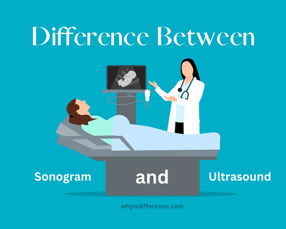
Sonogram and Ultrasound: Sonograms and ultrasound scans have become an everyday occurrence in hospitals today, commonly used to check fetuses during pregnancies for medical complications and ensure healthy fetal development. Sonography refers to another term for ultrasound scanning; both ultrasounds and sonographies refer to sound waves too high-pitched for humans to hear that can be used as medical exams to examine internal organs and fetuses whereas sonograms are images produced during an ultrasound exam.
What is an Ultrasound?
Ultrasound imaging is an innovative non-invasive diagnostic technology used by physicians and technicians alike, to gain insights into a patient’s internal structures and organs without having to make incisions or perform any procedures. Philips IE33 ultrasound machines allow doctors and technicians to observe these organs with no incisions required – an invaluable benefit that allows accurate diagnoses without surgery or procedures is needed. Sonography, commonly referred to as ultrasound imaging, often causes confusion.
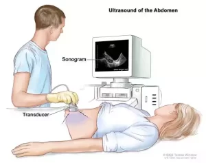
Ultrasound machines generate images by emitting high-frequency soundwaves from a transducer probe and using this energy to bounce off inner structures before returning back into their source transducer probe for processing. The angle and speed with which waves return to the probe indicate where objects are. This information is sent to a processor where algorithms are applied and computations completed before an image appears on a monitor screen. Ultrasound images are commonly used during pregnancy to check on development and growth; doctors also utilize ultrasound technology for diagnosing broken bones, cancerous tumors or chronic pain conditions.
What is a Sonogram?
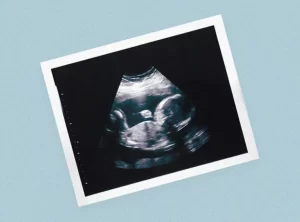
A sonogram is the result of all the data being fed into a processor. Sonograms can also be seen as another term for image, representing real-time images displayed on screen as well as any still photos captured during an exam or procedure. Both terms can easily be confused and used interchangeably – they both depend on each other for existence.
Uses of Ultrasound in Animal Kingdom
- Humans are visual species. Our experience of the world relies heavily on sight. By contrast, other animals rely on sonar or ultrasound waves to perceive their environment.
- Whales and Dolphins use ultrasonic sound waves to locate prey.
- Bats use ultrasound technology to locate their food source as bats have the ability to perceive frequencies between 100 and 200 kHz.
- Many insects such as tiger moths and beetles possess highly developed ultrasonic hearing, making their ultrasonic hearing superior in order to avoid being captured by bats.
- Dogs can hear frequencies between 18kHz and 22kHz.
- Fishes such as ray-finned species of fish have the ability to detect ultrasonic noises.
- ome species, such as Amolops enormous, utilize ultrasound waves as a form of communication.
- Grasshoppers & Mice – Both grasshoppers and mice use ultrasound waves to make mating calls.
Importance of Sonogram and Ultrasound in medical imaging
Sonograms and ultrasounds are essential medical imaging tools. They play an instrumental role in diagnosing and monitoring various medical conditions. This section will examine their roles within medical imaging.
Early identification and diagnosis:
- Sonogram, Obstetric sonograms are integral in monitoring fetal development, identifying anomalies, and determining due dates.
- Sonograms can assist in diagnosing conditions like ectopic pregnancy, multiple pregnancies, and placental abnormalities.
- These devices enable early diagnosis and treatment of malformations in fetuses.
Ultrasound Imaging:
- Cardiology and radiology specialties employ ultrasound imaging for early detection and diagnosis purposes.
- This test can identify abnormalities in organs such as the heart, liver, and kidneys.
- Ultrasound imaging can be used to diagnose conditions such as gallstones and cysts.
Safety and Non-Invasiveness:
- Sonogram, Sonograms are non-invasive and harmless to health; with no radiation exposure involved. Healthcare professionals as well as patients alike can use them.
- Due to their proven safety profile and ability to safeguard developing fetuses from potential danger, antiemetics are frequently prescribed during gestation.
- Ultrasound, Whilst radiography exposes patients to radiation, ultrasound does not cause this effect and thus offers non-invasive imaging options for diagnosing conditions.
This technique can be especially helpful for children or those allergic to contrast-enhanced images or who suffer from other medical issues.
Real-Time imaging and guided procedures:
Sonogram:
- Sonograms provide healthcare providers with real-time images of an expectant mother or its internal organs moving.
- CTG strips provide guidance and precise localization during procedures like amniocentesis and chorionic-villus sampling.
Ultrasound:
- Ultrasound-guided techniques are frequently employed for performing injections, biopsies, and aspiration procedures.
- Accurate needle placement allows for more successful procedures with decreased risks and success rates.
Cost-Effectiveness and Portability:
- These features make a product cost-effective and portable.
- Sonography equipment can easily be transported between healthcare settings such as clinics and hospitals.
- Sonograms offer an economical solution for screenings and follow-up exams.
Ultrasound:
- Ultrasound machines offer portable imaging for various imaging applications at an economical price point.
- These devices can be utilized at the point of care, in emergency situations, and in regions with limited resources to increase access to healthcare for populations that are underserved.
Sonogram and ultrasound imaging techniques have become indispensable tools in medical imaging, providing early detection, accurate diagnoses, and guided interventions for patient safety. Sonograms and ultrasounds have proven cost-effective due to non-invasiveness and real-time imaging capabilities; further revolutionizing modern healthcare by improving patient outcomes and decision-making capabilities.
Comparison Table:
| Sonogram | Ultrasound | |
|---|---|---|
| Definition | A sonogram refers to the image or visual representation produced using ultrasound technology. | Ultrasound is the imaging technique itself that uses high-frequency sound waves to create images of the body’s internal structures. |
| Function | Sonogram is the visual output or result of an ultrasound examination. It provides a detailed view of the internal structures of the body. | Ultrasound is the actual process of using sound waves to create images. It involves the transmission and reception of sound waves to generate real-time images. |
| Types of Images | Sonograms can be two-dimensional (2D) or three-dimensional (3D) images, allowing for detailed visualization of structures and their spatial relationships. | Ultrasound can produce various types of images, including 2D, 3D, and even four-dimensional (4D) images, which add a time component to the 3D view. |
| Usage | Sonograms are used to visualize specific areas of interest and provide detailed diagnostic information. They are often used to examine organs, tissues, and developing fetuses during pregnancy. | Ultrasound imaging is a broader term that encompasses various medical applications. It can be used for diagnostic purposes, monitoring conditions, guiding interventions, and assessing blood flow. |
| Terminology | The term “sonogram” is commonly used in reference to obstetric and gynecologic imaging, particularly during pregnancy. | “Ultrasound” is the general term used to describe the imaging technique across different medical specialties. |
| Focus | Sonogram focuses on the visual representation of anatomical structures and their characteristics, such as size, shape, and position. | Ultrasound focuses on the entire imaging process, including the generation, transmission, and reception of sound waves, as well as the interpretation of the acquired images. |
Ultrasound
- Ultrasonography, commonly referred to as a sonogram, is a vital imaging technology in many medical disciplines. Most notably it’s employed in obstetrics, diagnostic imaging and other fields where monitoring fetal growth in pregnancy or diagnosing medical conditions are common uses.
- Sonograms are created through high-frequency sound waves produced by a transducer. These sound waves travel throughout organs and tissues of the body, encountering various structures. A transducer detects when sound waves bounce back when encountering tissue boundaries such as between solid and fluid tissues – this detection allows it to convert these echoes to electrical signals which are then processed into images.
- Sonograms are an indispensable asset in obstetrics. They offer healthcare providers insight into fetus development and help identify abnormalities, assess the health of infants, provide anatomy information about the head, limbs, and heart size of babies, and also determine the gestational period and placenta location.
- Sonograms have an array of diagnostic uses outside obstetrics. Sonograms can help assess and visualize various organs of the body such as the pancreas and reproductive organs; as well as detect conditions like tumors, cysts, or stones as well as abnormalities in blood vessels – they’re even useful tools in guiding surgical procedures and interventions.
- Sonograms offer many advantages over other forms of imaging technology. Notably, sonograms do not utilize radioactivity, making them safe for both patients and healthcare providers alike. Their safety profile becomes especially crucial during gestation when they can monitor fetal development and health monitoring.
- Sonograms offer real-time images to healthcare professionals that enable them to see structures moving. This real-time imaging is particularly helpful during procedures like amniocentesis where precise guidance is required in extracting amniotic liquid for diagnostic use – sonograms provide real-time guidance that ensures accuracy while mitigating risks.
- Sonograms are essential tools in medical imaging, particularly obstetrics, as they allow healthcare providers to rapidly diagnose conditions and guide interventions. Sonograms have revolutionized healthcare with their noninvasiveness and real-time imaging capabilities; as well as having many uses.
Procedure and Equipment
- Ultrasound imaging (also referred to as ultrasonography) is widely utilized across numerous medical specialties for both diagnosis and monitoring purposes. Ultrasound uses high-frequency soundwaves to produce images of internal structures within the body which provides valuable insight for medical professionals.
- Transducers emit soundwaves into the body and collect their echos when they meet tissues or organs, then converts them to electrical signals which computers then use to generate real-time images on monitors.
- Diagnostic ultrasound imaging can offer unique insight into various body systems, particularly organs like liver, heart, kidneys and spleen. By visualizing these structures medical professionals can detect abnormalities quickly as well as assess organ functions to monitor disease progression and progression.
- Ultrasound imaging offers numerous advantages that make it suitable for patients of all ages and conditions – not least being non-invasive and using no ionizing radio waves like other imaging modalities such as X-rays or computed tomography scans (CT). As such, ultrasound offers safe and effective care options to everyone, including children and pregnant women.
- Ultrasound imaging has become an indispensable medical guidance tool. It assists doctors in pinpointing anatomical features and pinpointing needle placement for injections, biopsies, or aspiration procedures – real-time imaging provides increased accuracy and safety to reduce complications.
- Ultrasound imaging stands out for its diverse abilities to produce images in different forms, from two-dimensional (2D), three-dimensional (3D), and four-dimensional (4D). While two-dimensional (2D), imaging remains the most popular, ultrasound also generates three- and four-dimensional (4D) imagery which provides enhanced depth perception and visualization which allow a comprehensive assessment of anatomical structures as well as diagnosing complex medical conditions more quickly and accurately.
- Ultrasound imaging can be carried around and is relatively affordable compared to other forms of imaging. Because of its portability, ultrasound can be utilized in numerous healthcare settings including clinics and hospitals as well as emergency rooms and remote locations – making ultrasound widely popular as both affordable and accessible imaging option.
- Ultrasound imaging has completely revolutionized medical imaging. Thanks to its noninvasive nature, real-time capabilities and diverse applications it offers, ultrasound has become an indispensable tool for diagnosing and monitoring medical conditions. Furthermore, ultrasound provides vital data that medical professionals rely on every day.
- Users and Equipment for their Safety. Presented are some procedures and equipment necessary for safe operation of this system.
- Prep For Ultrasound Exam To prepare the patient for an ultrasound examination, position her for examination – in the case of obstetrical ultrasounds for example, this means having her lying on her back so as to expose all areas being examined – this may necessitate changing position or taking off clothing that covers areas being studied if necessary.
- When conducting an exam on any area that needs examination, use a clear gel. This enables soundwave transmission while maintaining proper skin contact.
- Once applied to the skin, a handheld transducer should be securely placed over an area covered with gel. A sonographer (healthcare provider) then maneuvers this transducer to capture images at various angles and perspectives; they may ask their patient to hold their breath briefly or apply gentle pressure in order to better visualize certain structures.
- The transducer emits sound waves as it travels over the skin. As its oscillators detect echos and convert them to electrical signals, an ultrasound machine uses these signals to produce real-timeSonographers analyze images live, assessing anatomy, structures, and any potential abnormalities they find. Still, images or video sequences may be captured to serve as documentation and analysis purposes.
- A transducer is an essential component of an ultrasound system, emitting sound waves and receiving echos to create images. Transducers come in various sizes and shapes for various examination purposes.
- Ultrasound Machine – An ultrasound machine is a device that processes electrical signals from transducers into real-time images displayed on a monitor. The control panel provides various settings to customize imaging parameters and a display screen for monitoring image displays.
- Displays live ultrasound images that allow sonographers to observe, assess, and capture video or images of anatomic structures in real time.
- An ultrasound machine is connected to a computer that processes its transducer data. With software capabilities allowing the manipulation of images, measurements, and documentation.
- Some ultrasound machines come equipped with built-in printers that enable instantaneous image printing and examination details to be stored digitally within the system, or transferred directly into an image archiving system for long-term storage.
Here are the steps and equipment necessary for performing a sonogram, though specific methods and devices used may depend on your examination type and settings.
Benefits and Limitations
Benefits:
- Non-Invasiveness – Sonograms and ultrasound imaging are non-invasive treatments, meaning no incisions need be made or instruments inserted into the body – making these diagnostic techniques safer and more comfortable for patients than their invasive counterparts.
- Ultrasound and sonography use sound waves rather than ionizing radiation, making them safer for patients, particularly pregnant women and young children who may be at higher risk due to any potential harmful radiation exposure.
- Real-Time Imaging, Sonograms and ultrasounds offer real-time imaging that allows healthcare professionals to monitor dynamic processes and moving structures within the body in real-time, which is especially beneficial when conducting procedures that require precise guidance or interventions requiring guidance.
- Sonograms and ultrasound images can be utilized to examine various body systems, such as the abdomen and pelvis, heart, blood vessels and musculoskeletal structures. Furthermore, sonograms serve as a valuable way of diagnosing medical conditions of various sorts.
- Cost-Effectiveness, Ultrasound machines tend to be more cost-effective than other imaging modalities such as computed tomography or magnetic resonance imaging, making ultrasound an accessible option for regular screenings, follow-up exams and point-of-care imaging.
- Ultrasound machines are lightweight and easily portable, making them suitable for use in hospitals, clinics, emergency rooms and remote or resource-limited locations.
Limitations:
- Operator Dependence: The quality and accuracy of ultrasound and sonogram images depend heavily on the skill and experience of the operator. To obtain optimal images, operators may need to have mastery over image optimization and transducer manipulation techniques.
- Limitations in Tissue Penetration: Ultrasound waves have limited penetration capabilities when applied to certain tissues such as air-filled structures or bone, making it more challenging to see deep structures and produce subpar image quality results.
- Image Interpretation: Interpreting sonograms and ultrasound images may be challenging in certain instances, such as when imaging obese individuals or those who contain significant amounts of abdominal gas. Furthermore, distinguishing between types of tissue or detecting subtle abnormalities may prove challenging; further imaging methods may be required for confirmation purposes.
- Operator Fatigue: Sonograms and ultrasonic examinations can be physically demanding, particularly when performing long scans on patients with complex anatomical conditions or long scanning times.
- Specific Applications: While sonograms and ultrasounds can provide many beneficial applications, some medical conditions require the assistance of other imaging modalities like MRI or CT for comprehensive evaluations. Sometimes ultrasound does not offer enough detail or visualization needed in these instances.
Clinical Applications
Sonogram and ultrasound images have many clinical applications across different medical specialties, from diagnosing to monitoring conditions to providing guidance during intervention. Here are a few applications you may encounter:
Obstetrics and Gynecology
- Obstetrics & Gynecology Sonograms play an integral part in monitoring fetal development during gestation. They can help determine gestational age as well as detect abnormalities.
- Ultrasound imaging can be used to diagnose conditions like ectopic pregnancy, multiple pregnancies and placental abnormalities.
- Gynecological ultrasound imaging helps evaluate reproductive organs like the uterus and ovaries to diagnose conditions like cysts or fibroids in their female hosts.
Cardiology:
- Ultrasound imaging, commonly referred to as echocardiography, provides essential analysis of heart structure and function.
- This test helps to diagnose heart conditions such as abnormal valve function or tumors in the heart.
- Echocardiography provides information on heart chamber dimensions, blood flow patterns and any abnormalities with heart muscle contraction.
Radiology:
- Whilst radiology, sonograms, and ultrasound imaging technologies are utilized for diagnosing different organs and systems within the human body, sonography and ultrasound images provide another tool.
- An abdominal ultrasound can be used to assess liver, gallbladder and kidney function as well as to examine other organs such as spleens, pancreas and blood vessels in the abdomen – helping diagnose conditions such as gallstones and liver cysts.
- Musculoskeletal Ultrasounds are used to assess joints, tendons, and ligaments. They assist with diagnosing tendonitis and bursitis as well as muscle tears, joint effusions and bursitis.
Vascular Medicine:
- Ultrasound imaging can be utilized in vascular medicine to analyze blood vessels and flow, detect abnormalities, and monitor health conditions.
- Doppler Ultrasound measures blood flow speed and direction to help diagnose conditions like deep vein thrombosis.
- The information gained here can also help guide procedures such as venous ablation and arterial interventions.
Emergency Medicine:
- Ultrasonography has become an invaluable asset in emergency care settings to rapidly diagnose critical conditions quickly.
- Ultrasound can be an invaluable way to assess trauma victims for internal bleeding, organ damage, and foreign bodies.
- Diagnostic imaging technologies aid in the identification and treatment of conditions like appendicitis and gallbladder inflammation.
Interventional Procedures:
- Ultrasound guidance is an essential component of many interventional procedures such as biopsies and aspirations.
- Precision aids the accuracy and success rate of these procedures while decreasing complications.
- Here are a few clinical applications of sonograms and ultrasound imaging. Sonograms and ultrasounds have become indispensable tool in many medical specialties thanks to their non-invasive imaging capabilities and real-time image delivery, providing healthcare providers with invaluable diagnostic information on an array of conditions.
Cost and Accessibility
Cost:
- Equipment, The cost of ultrasound equipment varies based on its manufacturer, model and features. While ultrasound machines tend to be more affordable than other imaging modalities such as MRI or CT scanners, high-end systems or those specialized for specific tasks may have higher expenses associated with them.
- Maintenance and Upgrades. Regular servicing and maintenance is essential to the longevity and optimal performance of ultrasound equipment, including any upgrades or software updates that might occur over time. Include the costs for maintenance along with any upgrades or software updates in your total price quote.
- Healthcare professionals require training on ultrasound equipment in order to use it efficiently, such as sonographers and radiologists. Training programs and certification costs may become burdensome for healthcare institutions and individuals.
- Consumables, When considering the cost of consumables such as probe covers and ultrasound gel, which are required for sonograms and ultrasonography exams, take an account of all related expenses.
Accessibility:
- Healthcare Settings Ultrasound imaging can be utilized in various healthcare settings such as hospitals, clinics, and private practices. Mobile imaging units are also available and allow ultrasound imaging to reach remote areas or underserved communities more readily than before.
- Ultrasound Machines in Healthcare Settings, Ultrasound machines have become an invaluable asset to emergency rooms, intensive-care units, and other ambulatory settings across the United States, enabling quick assessment and decision-making at the point of care. Their accessibility enables rapid imaging that facilitates immediate imaging for quick decision-making and rapid assessment.
- Ultrasound imaging is considered more cost-effective than other forms of imaging technology, making it suitable for routine screenings, follow-up exams and chronic condition monitoring.
- Non-Invasiveness – Ultrasound imaging’s noninvasive nature and accessibility make an ultrasound examination accessible for patients of all ages, including children and pregnant women, without experiencing significant discomfort or radiation exposure.
- Ultrasound Is Ideal in Resource-Limited Settings, Ultrasound is an invaluable imaging solution in resource-limited settings such as developing countries and areas without access to advanced imaging technology. Portable ultrasound machines enable healthcare providers to provide essential diagnostic services even in challenging conditions.
Sonogram and ultrasound imaging costs and accessibility vary based on factors like geographical location, healthcare infrastructure, insurance coverage and availability of skilled operators. To ensure equitable access for all, healthcare institutions and policymakers must consider cost-effective strategies and training programs as well as appropriate resource allocation to guarantee equitable treatment for everyone.
Safety and Risks
Sonograms and ultrasound imaging are widely viewed as safe diagnostic tools with limited risks associated with them. Here is a brief overview of potential safety concerns and risks.
- Safety, Sonograms, and ultrasound images use sound waves instead of radioactive radiation to produce images, providing less radiation exposure – particularly beneficial for children and pregnant women who may be vulnerable.
- Sonograms and ultrasound imaging do not involve surgery, needles or contrast agents – and are generally well tolerated without causing pain or discomfort to patients.
- Real-Time Imagery, Sonograms and ultrasounds offer healthcare professionals real-time images, allowing them to monitor body structures moving inside a person’s body in real-time and assess dynamic processes as they occur – providing immediate assessment and guidance, which reduces further interventions needed in later treatments.
- Lack of Long-Term Risks, Presently there are no long-term known risks associated with sonograms and ultrasound imaging performed by trained healthcare professionals using appropriate equipment.
- Operator Skill and Interpretation – The accuracy and quality of sonogram images depend heavily on an operator’s skills, experience, and interpretation when acquiring and interpreting images. To obtain high-quality images it is imperative that an operator is proficient with optimizing and manipulating transducers.
- False Positives or False Negatives. Although sonograms and ultrasound imaging can detect certain abnormalities, they may occasionally produce false-positives or false-negatives that compromise accurate diagnosis. For an accurate assessment, additional imaging or clinical correlation may be required for more accurate assessment.
- Ultrasound Imaging for Invasive Procedures – Ultrasound imaging is sometimes used to assist with more invasive procedures, like biopsy or aspiration. Such procedures may carry risks including infection and bleeding – these will depend on which one you decide on.
- Limitation in Tissue Penetration, Ultrasound waves have a limited ability to penetrate certain types of tissues such as bone or air-filled structures, leading to an impaired view of deeper structures that could hamper diagnostic accuracy.
- Tissue heating can occur occasionally with ultrasound machines and protocols; however, modern ultrasound machines and guidelines include safety features to minimize this risk.
Sonograms and ultrasound imaging typically outweigh their risks due to being noninvasive and real time capable, as well as not emitting ionizing radio waves. Healthcare professionals and sonographers follow established protocols and safety guides when performing sonograms and ultrasound imaging for patient safety purposes.
Future Developments
Sonograms and ultrasound imaging will continue to develop due to ongoing research and technological innovation, as evidenced by some promising developments. Here are some such developments.
- Enhancing Image Quality, Ultrasonic technology advancements aim to improve image quality and resolution. Transducers with higher frequencies, advanced signal processing algorithms and more effective image reconstruction techniques will enhance ultrasound images’ clarity and detail, leading to more accurate diagnosis.
- Three-Dimensional Imaging (3D) and 4-Dimensional Imaging (4D), Four-dimensional and three-dimensional ultrasound techniques have become increasingly popular, providing volumetric imaging as well as real-time visualization of moving structures within the body for more comprehensive assessments of complex anatomical processes and dynamic structures.
- Contrast-Enhanced Ultrasound (CEUS) Imaging, Contrast agents are frequently used in ultrasound imaging to improve the visibility of blood flow and to better characterize certain tissues or lesions. Contrast-enhanced ultrasound (CEUS), is particularly helpful for diagnosing liver lesions as well as differentiating between benign and cancerous masses.
- Elastography Utilizing ultrasound waves, Elastography is used to evaluate tissue stiffness. This test is particularly helpful in evaluating thyroid, breast or liver stiffness – and can even assist in diagnosing potential tumors and other fibrotic conditions.
- Point-of-Care and Handheld Devices, With the introduction and proliferation of portable and hand-held ultrasound devices, point-of-care imaging is now possible in various environments such as emergency care, field medical, and environments with limited resources. These devices provide immediate diagnostic information while increasing access to ultrasound imaging services.
- Artificial Intelligence and Machine Learning, Artificial intelligence and machine learning techniques are being integrated into ultrasound imaging for image analysis, automation and decision support. AI algorithms have proven their worth in improving image interpretation as well as automating measurements – increasing efficiency and accuracy during ultrasound examinations.
- Fusion Imaging, Combining ultrasound with other imaging modalities like MRI or CT can offer complementary information to increase diagnostic accuracy, providing for increased visualization of anatomical structures as well as precise targeting and treatment planning. This technique is known as Fusion Imaging.
- Ultrasound techniques are increasingly being explored for therapeutic applications. High-intensity focus ultrasound (HIFU) has proven particularly successful at non-invasively treating tumors, dispensing targeted drugs, and disrupting blood clots – these applications promise precise yet minimally-invasive interventions.
Future developments could significantly advance sonograms and ultrasound imaging technology. Such advancement could improve diagnostic accuracy, patient outcomes and accessibility to ultrasound imaging across healthcare settings.
Conclusion
While ultrasounds and sonograms share the same fundamental concept that uses sound waves to aid in imaging, they have distinct functions within the field of medicine. Sonograms are focused on observing the development of a fetus during pregnancy, and fostering the relationship between parents and the unborn baby. Contrary to ultrasounds, ultrasounds offer greater scope that allows healthcare professionals to detect and monitor various medical conditions throughout different body systems.
Understanding the distinctions of these different imaging methods is crucial in recognizing the unique contribution they make to the field of healthcare. As medical technology advances sonograms and ultrasounds are certain to play an important role to shape the direction of medical research by offering patients better treatment and enhanced diagnostic capabilities.